



A Real Case from Orthopedic Practice
Every week in orthopedic trauma practice, patients present with serious knee injuries resulting from road traffic accidents, falls from height, and sports-related trauma. One such injury is a tibial plateau fracture, which affects the upper end of the tibia (shin bone) and involves the knee joint.
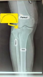
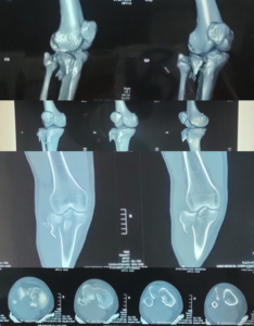
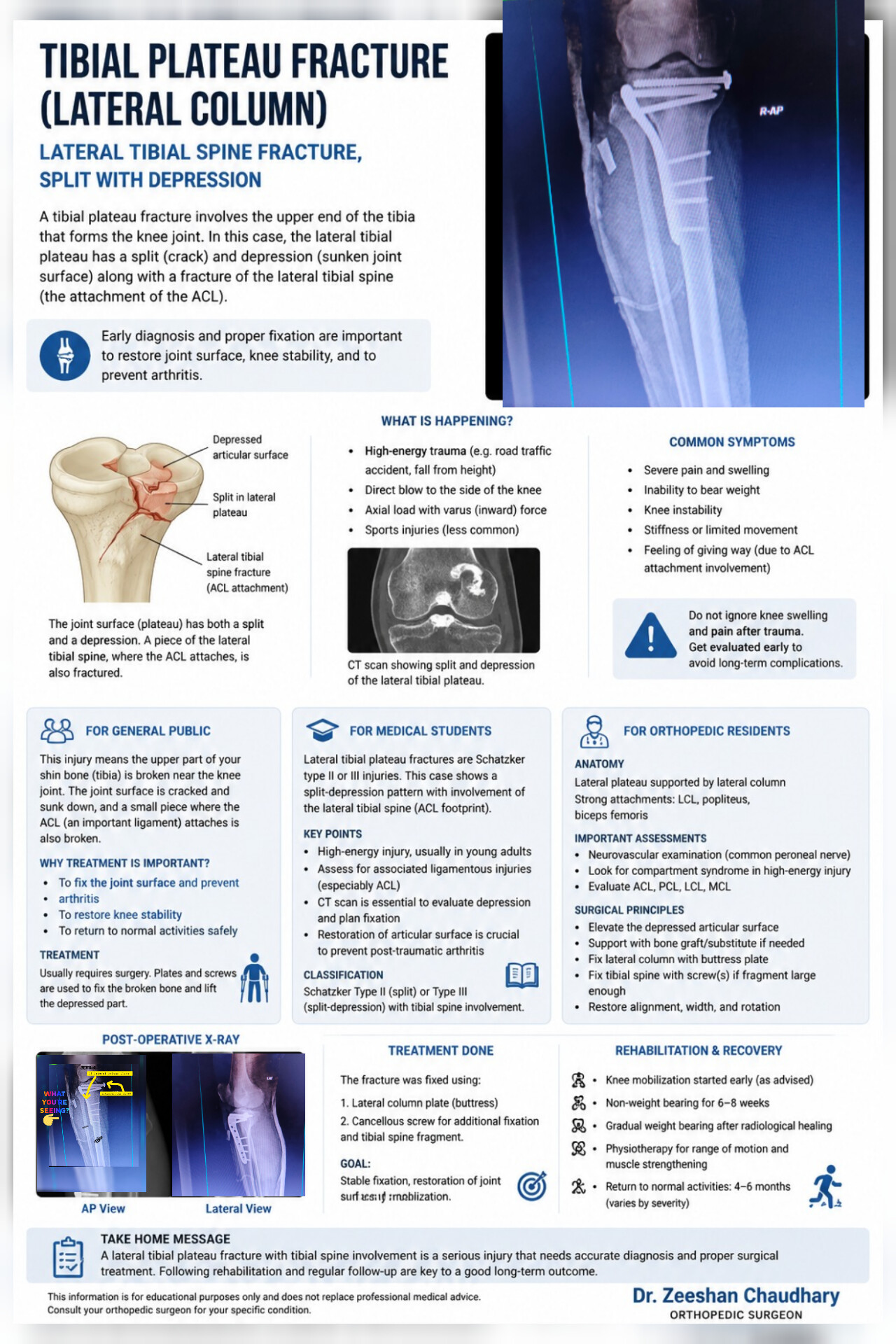
This real case demonstrates a lateral tibial plateau split-depression fracture associated with a lateral tibial spine fracture, successfully treated using a lateral column buttress plate and cancellous screw fixation.
Although this injury is commonly seen in orthopedic trauma, many patients are unaware of its seriousness. If left untreated, it can lead to chronic pain, knee instability, stiffness, and early arthritis.
What Is a Tibial Plateau Fracture?
The tibial plateau forms the lower half of the knee joint. It is the smooth upper surface of the tibia where the femur rests during standing, walking, and running.
When a high-energy force strikes the knee, the articular surface may crack, split, or become depressed. Instead of remaining smooth, the joint surface sinks downward, creating an uneven weight-bearing surface.
In this patient, the injury included:
- Split fracture of the lateral tibial plateau
- Depression of the joint surface
- Fracture of the lateral tibial spine (ACL attachment)
Because the fracture involved the knee joint itself, accurate reduction and stable fixation were essential to restore normal knee function.
How Do These Injuries Usually Occur?
The most common causes include:
- Road traffic accidents
- Motorcycle injuries
- Falls from height
- Sports injuries
- Direct impact to the side of the knee
Why Is This Injury Important?
Unlike simple fractures, a tibial plateau fracture involves the joint surface.
If the depressed fragment is not elevated back to its normal position, the knee joint no longer remains smooth.
This can result in:
- Early osteoarthritis
- Persistent knee pain
- Loss of knee motion
- Difficulty walking
- Knee instability
- Poor long-term knee function
For this reason, accurate restoration of the joint surface is one of the primary goals of treatment.
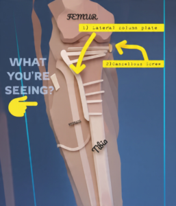
Understanding the Postoperative X-ray
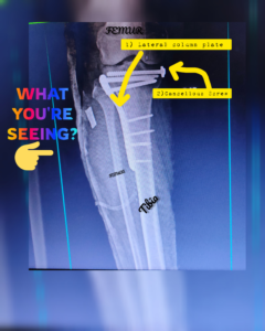
Lateral Column Buttress Plate
A specially designed buttress plate was applied along the lateral side of the tibia.
Its purpose is to:
- Support the fractured lateral column
- Prevent collapse of the elevated joint surface
- Maintain alignment throughout healing
Cancellous Screw Fixation
A cancellous screw was inserted to provide additional compression and stabilize the articular fragments, including the tibial spine fragment.
Together, these implants restore the normal anatomy while allowing the fracture to heal in the correct position.
Surgical Treatment
The goal of surgery is not simply to repair a broken bone—it is to reconstruct the knee joint and restore its normal function.
The procedure generally includes:
- Elevation of the depressed articular surface
- Anatomical reduction of the fracture
- Stabilization using a lateral buttress plate
- Fixation of associated fragments with cancellous screws
- Confirmation of alignment using intraoperative imaging
Stable fixation allows early knee movement while protecting the repaired joint during healing.
Recovery After Surgery
Recovery depends on the severity of the fracture and the patient’s commitment to rehabilitation.
Most rehabilitation protocols include:
- Early knee range-of-motion exercises
- Non-weight bearing for approximately 6–8 weeks
- Gradual progression to partial and full weight bearing after radiological healing
- Physiotherapy to restore muscle strength and knee function
Most patients continue improving for several months after surgery.
Key Takeaway
Think of your knee joint like the smooth surface of a road.
If a section of the road collapses, every vehicle passing over it experiences an uneven ride.
The same principle applies inside the knee.
If the depressed joint surface is not restored, every step places abnormal pressure on the cartilage, increasing the risk of arthritis and chronic pain.
Modern orthopedic surgery aims to restore this surface as accurately as possible so patients can return to normal daily activities with the best possible long-term outcome.
Orthopedic & Trauma Surgeon
Conclusion
A tibial plateau fracture is much more than a broken bone—it is a joint injury.
Successful treatment depends on:
- Restoring the smooth articular surface
- Maintaining knee stability
- Achieving stable fixation
- Beginning appropriate rehabilitation
With timely surgery and dedicated physiotherapy, many patients regain excellent knee function and return to their normal daily activities.
Frequently Asked Questions
Is a tibial plateau fracture a serious injury?
Yes. Because it involves the knee joint surface, accurate treatment is essential to restore normal knee function and reduce the risk of long-term complications.
Does every tibial plateau fracture require surgery?
No. Treatment depends on the fracture pattern, displacement, joint involvement, and patient factors. Some fractures may be managed without surgery, while others require surgical fixation.
How long does recovery take?
Recovery varies depending on the severity of the injury and rehabilitation progress. Most patients require several months for optimal recovery.
When can I start walking after surgery?
Most patients remain non-weight bearing for approximately 6–8 weeks, followed by gradual progression based on clinical and radiological healing, as advised by the treating orthopedic surgeon.
About the Author
Orthopedic & Trauma Surgeon
Department of Orthopaedic & Spine Surgery
Mayo Hospital Lahore
Dedicated to providing evidence-based orthopedic care with a focus on trauma surgery, joint preservation, fracture management, and patient-centered rehabilitation.
Clinical Note: This case has been shared for educational purposes. Patient privacy has been protected, and any clinical images are used with appropriate consent and anonymisation where applicable.
Leave a Reply