



A Case From the Surgical Diaries
A fracture does not always heal simply because the bone has been fixed with a plate or nail.
Sometimes, despite surgery, the fracture fails to progress toward union. This can result from problems with biology, stability, infection, patient factors, or a combination of these factors.
This is called nonunion.
In this article, I will explain nonunion in simple terms and discuss a young patient with nonunion of both the radius and ulna after previous fracture fixation.
What Is Nonunion?
Bone healing is a gradual biological process. After a fracture, the body forms new tissue and bone around the fracture site and progressively restores continuity and strength.
A fracture is generally considered a nonunion when at least 9 months have passed since the injury, and there have been no visible progressive signs of healing for approximately 3 consecutive months.
Nonunion is therefore not simply “a fracture that has not healed yet.”
It means that the normal healing process has effectively stalled.
What Is Delayed Union?
Delayed union means that a fracture is taking longer than expected to heal.
There is no single time period that applies to every fracture. Healing depends on:
- The bone involved
- Fracture pattern
- Soft-tissue injury
- Blood supply
- Patient factors
- Stability of fixation
- Presence of infection
A delayed union can progress to nonunion if the underlying problem is not identified and corrected.
Early recognition of delayed healing may provide an opportunity to intervene before established nonunion develops.
Why Does Nonunion Occur?
Nonunion is usually caused by one or more problems involving biology or mechanics.
1. Biological Factors
Local Factors
These may include:
- Severe soft-tissue injury
- Loss of bone or soft tissue
- Poor local blood supply
- Infection
- Previous surgery
- Radiation
- Extensive periosteal damage
Systemic Factors
The patient’s overall health can also influence bone healing.
Important factors include:
- Advanced age
- Malnutrition
- Smoking or nicotine exposure
- Chronic anemia
- Diabetes and other chronic diseases
- Metabolic or endocrine abnormalities
- Vitamin D deficiency
- Certain medications
- Poor immune status
2. Mechanical Factors
Even when the biology is adequate, a fracture may fail to unite if the mechanical environment is inappropriate.
Examples include:
- Poor fracture reduction
- Excessive movement at the fracture site
- Inadequate fixation
- Inappropriate implant selection
- Implant loosening or failure
- An unstable construct
Bone healing requires both appropriate biology and appropriate mechanical stability.
Classification of Nonunion
Nonunion can be classified in several ways.
According to Location
- Epiphyseal
- Metaphyseal
- Diaphyseal
According to Infection
- Septic
- Aseptic
According to Radiographic Appearance
- Hypertrophic
- Oligotrophic
- Atrophic
The appearance of the fracture site can provide clues about the underlying problem.
Pseudoarthrosis
In established cases, a false joint or pseudoarthrosis may develop at the nonunion site, with abnormal movement between the bone fragments.
What Symptoms Suggest Nonunion?
Patients may present with:
- Persistent pain at the fracture site
- Pain with loading or activity
- Abnormal movement at the fracture site
- Persistent tenderness
- Deformity
- Reduced function
- Failure to regain normal limb use
Some patients may have surprisingly little pain, particularly if the nonunion has become chronic.
Therefore, symptoms must always be interpreted together with clinical examination and imaging.
How Do We Evaluate a Suspected Nonunion?
A detailed history is essential.
We need to understand:
- How the original injury occurred
- The original fracture pattern
- Previous treatment
- Previous operations
- Previous infections
- Immobilization and rehabilitation
- Nutritional status
- Smoking or nicotine use
- Medication history
- NSAID exposure
- Previous medical illnesses
Physical Examination
A complete examination should include:
- Local tenderness
- Abnormal mobility
- Deformity
- Limb alignment
- Soft-tissue condition
- Neurovascular examination
- Function of the affected limb
Investigations
Imaging
X-rays remain fundamental in evaluating fracture healing.
Depending on the case, CT may be useful to assess:
- Persistent fracture lines
- Bone bridging
- Alignment
- Union in complex anatomical regions
Laboratory Investigations
When nonunion is suspected, particularly before revision surgery, investigations may include:
- CBC
- ESR
- CRP
- Serum albumin
- Total protein
- Vitamin D
- Calcium and phosphate when indicated
- PTH when indicated
- Thyroid function when clinically indicated
The exact workup should be individualized.
Always Consider Infection
An apparently “aseptic” nonunion may occasionally have an occult infection.
Therefore, infection should be actively considered and appropriately investigated, particularly before revision surgery.
A Case From the Surgical Diaries
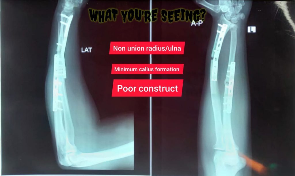
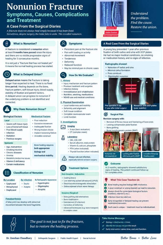
A young male patient presented approximately one year after previous fixation of fractures of the radius and ulna.
He had:
- No significant comorbidities
- No history of smoking
- No relevant medication history
- No obvious clinical evidence of infection
Previous fixation had been performed using DCPs.
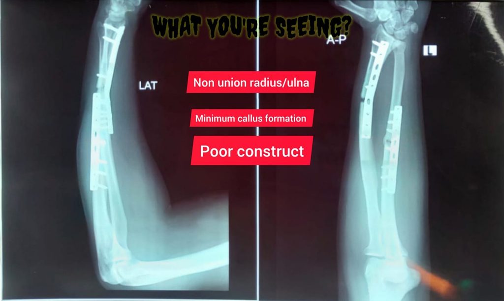
Radiographs demonstrated persistent nonunion of both the radius and ulna, with limited callus formation and an inadequate mechanical construct.
The overall picture was consistent with an oligotrophic nonunion associated with inadequate mechanical stability.
The Problem Was Not Simply “A Broken Bone That Had Not Healed”
In this patient, we had to consider two major components:
Biology
Was there sufficient biological activity to support healing?
Mechanics
Was the fracture adequately stabilized?
A fracture requires an appropriate biological environment and an appropriate mechanical environment.
If either is significantly compromised, union may fail.
Surgical Management
The patient was planned for revision surgery.
The principles of treatment included:
1. Preparation of the Nonunion Site
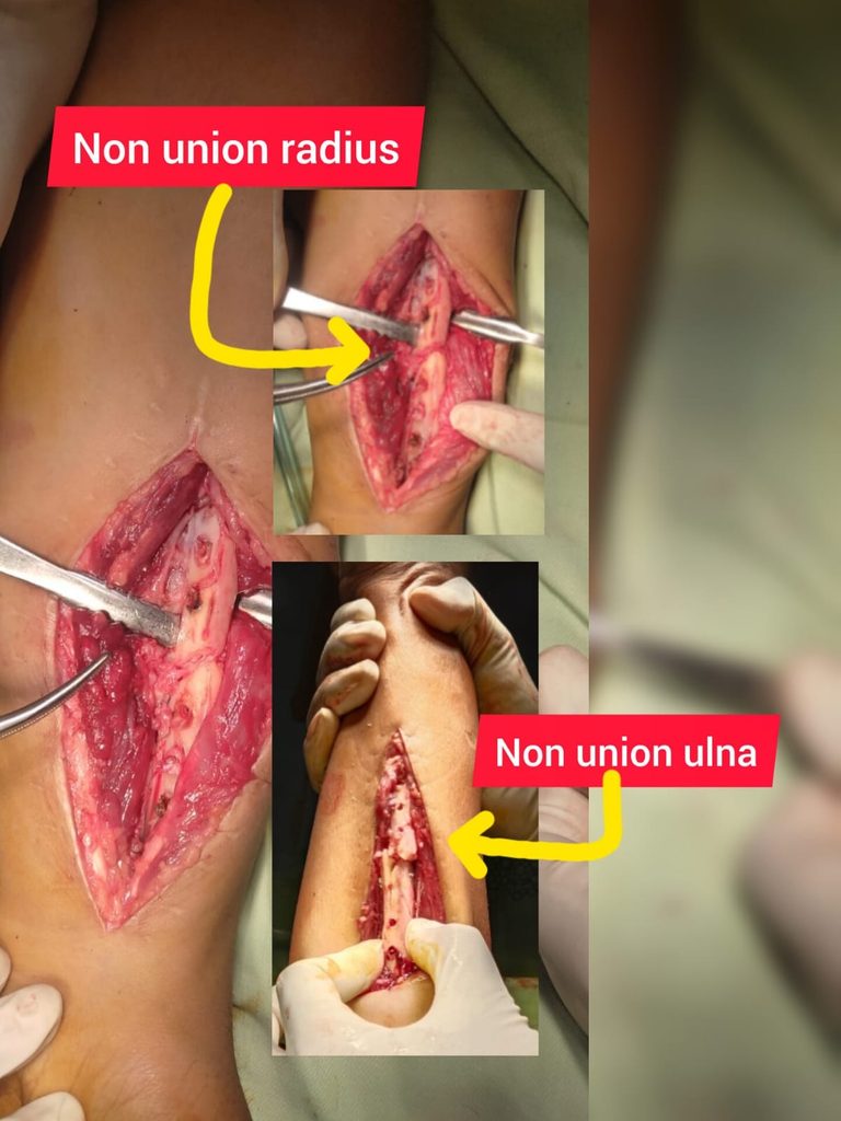
During surgery, the nonunion site was exposed.
Fibrous tissue was removed and the bone ends were freshened to create a biologically active environment.
2. Restoration of Stability
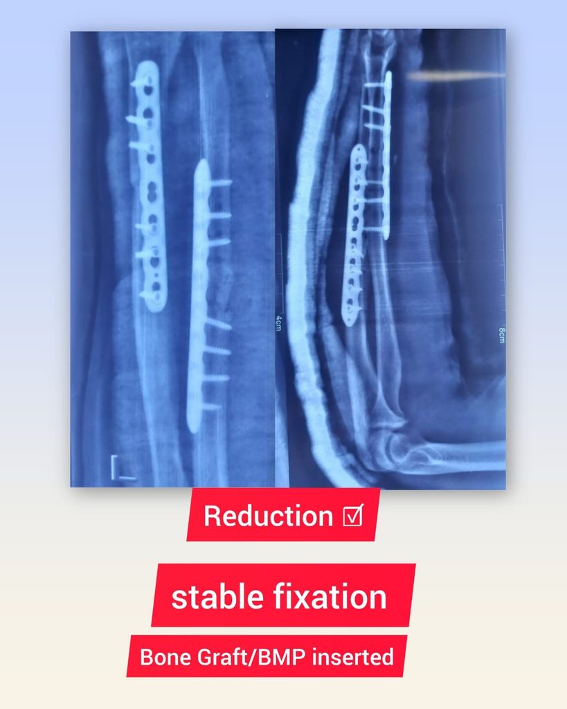
The previous construct was revised, and stable fixation was achieved using locking compression plates.
The aim was to obtain:
- Appropriate alignment
- Adequate stability
- Good fixation
- Preservation of viable biology
3. Bone Grafting
Bone graft was used to improve the biological environment at the nonunion site.
4. Biological Augmentation
A bone morphogenetic protein (BMP) was used as an adjunct according to the surgical plan and indication.
The specific use of biologic agents should always be individualized, considering evidence, indication, availability, regulatory considerations, and potential risks.
Follow-Up
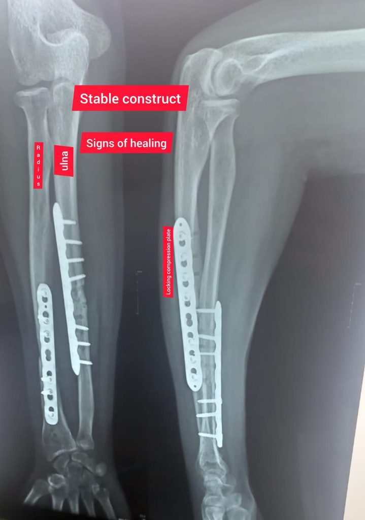
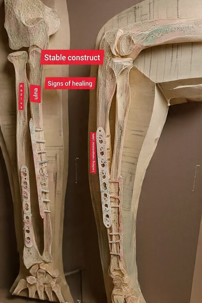
At approximately 3 months after revision surgery, follow-up radiographs demonstrated satisfactory progression toward union with evidence of healing.
The patient continued to be monitored clinically and radiologically.
What Does This Case Teach Us?
1. Bone healing requires biology AND mechanics.
A technically placed implant does not guarantee union.
2. A poor mechanical environment can contribute to nonunion.
Inadequate reduction or insufficient stability can prevent progression of healing.
3. Always look for correctable biological factors.
Nutrition, vitamin D status, smoking, infection, metabolic disease and other systemic factors should not be overlooked.
4. Infection must be considered.
Not every nonunion is sterile.
5. Early recognition matters.
Recognizing delayed healing and addressing the underlying problem may prevent progression to established nonunion.
6. Nonunion treatment is individualized.
There is no single operation that works for every nonunion. Treatment depends on:
Biology + Mechanics + Infection status + Bone loss + Patient factors + Location of fracture.
Treatment Options for Nonunion
Treatment can range from relatively simple measures to complex revision surgery.
Non-Invasive or Adjunctive Options
Depending on the individual case, these may include:
- Appropriate immobilization
- Activity modification
- Nutritional optimization
- Correction of metabolic abnormalities
- Low-intensity pulsed ultrasound
- Electrical or electromagnetic stimulation
- Extracorporeal shock-wave therapy in selected cases
Surgical Treatment
Depending on the cause, surgery may involve:
- Revision fixation
- Bone grafting
- Bone graft substitutes
- Debridement
- Correction of deformity
- Treatment of infection
- Bone transport or other reconstruction techniques in major bone defects
- Biological augmentation in selected cases
The key principle is not simply to “add bone graft.”
The cause of the nonunion must first be identified.
Final Take-Home Message
A nonunion is not merely a fracture that has taken a long time to heal.
It is a failure of the healing process that requires systematic evaluation.
When faced with a nonunion, ask:
Is the biology adequate?
Is the mechanical environment adequate?
Is there an infection?
Is there a correctable systemic problem?
The successful treatment of nonunion depends on identifying and correcting the underlying problem rather than simply repeating the original treatment.
Early recognition of delayed union, appropriate investigation, and timely intervention may prevent a difficult nonunion from becoming an even more complex problem.
From the Surgical Diaries
Every nonunion has a story.
Behind every radiograph is a patient, a previous treatment, a biological environment, a mechanical construct—and often an opportunity to understand why healing failed.
This case is presented for educational purposes. Patient-identifying information has been omitted or modified to preserve confidentiality.
Leave a Reply