


A Simple Guide for Patients, Medical Students, and Anyone Interested in Orthopedics
Every day in orthopedic practice, we see patients with fractures resulting from falls, road traffic accidents, sports injuries, and osteoporosis. One question patients often ask is, “Why did my bone break this way?” or “Why does healing take longer in some fractures?”
The answer lies in the fascinating structure of our bones.
Understanding how bones are built helps patients understand their injuries and helps medical students appreciate the principles behind fracture healing and fixation.
Bones Come in Different Shapes
Not all bones in the body are the same. Their shape and structure are designed according to their function.
Long Bones
These are the bones most commonly involved in fractures seen in orthopedic clinics.
Examples include:
- Femur (thigh bone)
- Tibia (shin bone)
- Humerus (upper arm bone)
- Radius and ulna (forearm bones)
Daily Practice Example
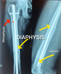
A young motorcyclist involved in a road traffic accident may sustain a fracture of the femur. Understanding the anatomy of long bones helps surgeons plan treatment using plates, screws, or intramedullary nails.
Flat Bones
These bones provide protection and support.
Examples include:
- Skull
- Pelvis
- Scapula (shoulder blade)
Daily Practice Example
Pelvic fractures are commonly seen after high-energy trauma and require a completely different approach compared to long bone fractures.
Inside Every Bone: Two Different Types
Although bones appear solid, they actually contain two different structures.
Cortical Bone – The Strong Outer Shell
Cortical bone makes up approximately 80% of the skeleton.
Characteristics:
- Dense and strong
- Provides mechanical support
- Resists bending and twisting forces
- Heals more slowly because of lower metabolic activity
Daily Practice Example
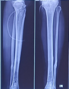
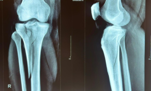
A tibial shaft fracture usually involves thick cortical bone and often requires months to achieve complete healing.
Cancellous Bone – The Spongy Inner Bone
Also called trabecular bone.
Characteristics:
- Lightweight
- Highly vascular
- More elastic
- Faster healing due to higher metabolic activity
Daily Practice Example
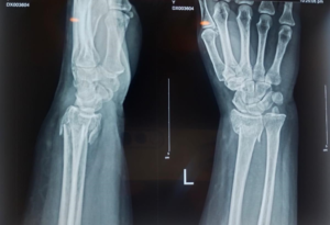
Distal radius fractures near the wrist and fractures around the knee often involve cancellous bone, which usually heals faster than shaft fracture.

Anatomy of a Long Bone
Long bones have three important regions.
Diaphysis (Shaft)
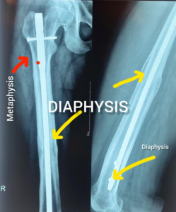
The central portion made mainly of strong cortical bone.
Common Clinical Example
Mid-shaft femur fractures in road traffic accidents.
Metaphysis
The transition zone between the shaft and the ends of the bone.
Contains abundant cancellous bone and excellent blood supply.
Common Clinical Example
Distal radius fractures and proximal tibial fractures.
Epiphysis
The ends of bones that form joints.
Covered by cartilage and responsible for smooth movement.
Common Clinical Example
Fractures involving the knee, shoulder, or ankle joints.
These injuries require accurate reduction to prevent arthritis later in life.
How Bones Heal: Woven Bone and Lamellar Bone
After a fracture, the body first produces immature bone called woven bone.
Woven Bone
Characteristics:
- Forms rapidly after injury
- Weak and disorganized
- Temporary structure
Daily Practice Example
Early callus seen on X-rays six weeks after fracture fixation.
Lamellar Bone
Over time, woven bone remodels into mature lamellar bone.
Characteristics:
- Organized structure
- Strong and stress oriented
- Permanent healthy bone
Daily Practice Example
Patients often ask, “Doctor, my fracture is united. Why does the X-ray continue changing for months?”
The answer is that remodeling continues long after the fracture has healed.
Why Osteoporosis Causes Fragile Bones
Healthy bone contains a balance between cortical and cancellous components.
In osteoporosis:
- Bone becomes porous.
- Trabeculae become thin.
- Bone strength decreases.
- Fractures occur even after minor falls.
Common Cases Seen Daily
- Hip fractures in elderly patients
- Wrist fractures after simple falls
- Vertebral compression fractures
Why Understanding Bone Structure Matters
Knowing how bones are structured helps orthopedic surgeons decide:
- Whether surgery is needed
- Which implant to use
- Why some fractures heal faster than others
- Why osteoporosis increases fracture risk
- How rehabilitation should be planned
For medical students, understanding cortical bone, cancellous bone, woven bone, and lamellar bone forms the foundation of fracture management and orthopedic surgery.
Key Takeaway
Bones are not simply rigid structures. They are living tissues with different regions and microscopic architectures designed to provide strength, flexibility, and healing potential. The same principles that explain normal bone anatomy also explain why fractures occur, how they heal, and why some patients recover faster than others.
Clinical Note: This case has been shared for educational purposes. Patient privacy has been protected, and any clinical images are used with appropriate consent and anonymisation where applicable.
Leave a Reply